Right right,
Here is the brooch design sheet:
http://s786.photobucket.com/albums/yy148/DesignBot/Drawings/?action=view¤t=ohfuck.png
The reason why the file name is ohfuck is that I did it waaaaaay too big in photoshop, so when I resize it smaller the quality is affected, now I don't know how this will affect the printouts, but seeing as we have to give in digital copies aswell the size shouldn't matter there. I was wondering if a copyshop would be able to sort it, I will have to go to one anyway as I don't have an A3 printer.
Do you think I should re-do them? Seeing as the address we have to have it to on Tues is in Imperial, I might take a bit more time and post it in by hand, is that even possible do you think?
Could you possibly write a bit of blurb/explanation as to the cells that we could include with the products? It would be really helpful in proving what we are trying to show scientifically.
What do you think of the designs so far? I have all the stages saved so I can play about with the whole thing. I used the first name you mentioned for our collection as I liked it but can change :)
Saturday, 29 August 2009


Thursday, 27 August 2009
Basics! Jewellery designs.
Made some basic images with prints that can be made up into jewellery designs.
Here they are:
http://s786.photobucket.com/albums/yy148/DesignBot/Drawings/
I am making up designs for the collection, will include the brooches we have talked about, the earrings pictured in the last update and necklaces based on the bipolar cell necklace idea.
How does this sound? :)
Here they are:
http://s786.photobucket.com/albums/yy148/DesignBot/Drawings/
I am making up designs for the collection, will include the brooches we have talked about, the earrings pictured in the last update and necklaces based on the bipolar cell necklace idea.
How does this sound? :)
Monday, 24 August 2009
what do retinal stem cells look like - good question...
3rd pic down on this page http://www1.imperial.ac.uk/medicine/about/divisions/neuro/npmdepts/cmn/cmnresearch/cmnplasticity/
And here http://neuromics.blogspot.com/2009/08/stemez-np1-neural-progenitors-now.html
Basically round, amorphous. They are not usually shown - they way you demonstrate that something is a stem cell is to make it generate lots of offspring, in a dish or transplanted into a retina. Stem cells are not visually distinctive, so there are more pictures of the distinct offspring.
And here http://neuromics.blogspot.com/2009/08/stemez-np1-neural-progenitors-now.html
Basically round, amorphous. They are not usually shown - they way you demonstrate that something is a stem cell is to make it generate lots of offspring, in a dish or transplanted into a retina. Stem cells are not visually distinctive, so there are more pictures of the distinct offspring.
Saturday, 22 August 2009
Tuesday, 18 August 2009
Muller cells
The glial cell type produced by retinal stem cells. A relatively unspecialised cell compared to the retinal neurons - no elaborate tree-like dendrites, etc. This cell type may be able to dedifferentiate back in to a retinal stem cell under certain conditions. Whether this happen in adults is controversial.
This group had an interesting paper - Muller cells act to refract light, helping to channel it down through the retina to photoreceptors and preventing distortion. Simpler explanation here
Picture here.
Apart from this, they have the usual glial cell function of support and nutrient supply to neurons. That cute knife-and-fork graphic can be used - if you decide to use prints. They might get a little confusing in the overlay, but for separate neuron cut-outs...
Images
Golgi (black)stained pics
and here
Fluorescent pics here
And here
This group had an interesting paper - Muller cells act to refract light, helping to channel it down through the retina to photoreceptors and preventing distortion. Simpler explanation here
Picture here.

Apart from this, they have the usual glial cell function of support and nutrient supply to neurons. That cute knife-and-fork graphic can be used - if you decide to use prints. They might get a little confusing in the overlay, but for separate neuron cut-outs...
Images
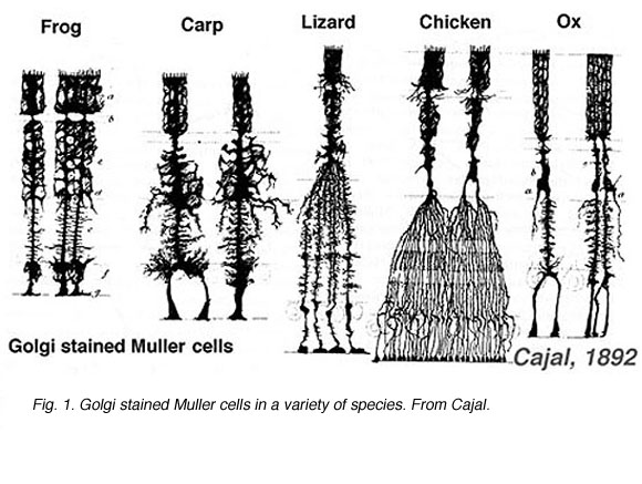
Golgi (black)stained pics

and here
Fluorescent pics here
And here
Monday, 17 August 2009
Retinal Neuron pictures
Amacrine cells
http://webvision.med.utah.edu/amacrines1.html
This one looks like the best source site for all cell types in the retina, which reminds me I need to talk about Muller cells next.
http://www.retinalmicroscopy.com/
The internet is a beautiful, beautiful thing to give us all these redunkulously specialised resources. If I ever left Neuroscience, it would be for computer programming.
http://webvision.med.utah.edu/amacrines1.html
This one looks like the best source site for all cell types in the retina, which reminds me I need to talk about Muller cells next.
http://www.retinalmicroscopy.com/
The internet is a beautiful, beautiful thing to give us all these redunkulously specialised resources. If I ever left Neuroscience, it would be for computer programming.
Retinal neurons and relationships, part 2
Hey, just thought of a design name. Retinal Images, anyone? See Me. The Shapes Behind Your Eyes. A Vision In Aluminium. Sight Unseen.
Stop me, please.
Anyway, The bipolar cell is firing. Let's talk about its destinations: Amacrine cells and Ganglion cells.
Amacrine cells are another processing waystation, like Horizontal cells in the last post. Except that if Horizontal cells were analogous to pressing 'sharpen image' in photoshop, Amacrine cells are like tuning contrast, colour balance and intensity. They are also involved in the perception of moving objects. They are complex and not well understood, like tiny cellular James Joyces. There are 20+ subtypes that use several different neurotransmitters. The do not have axons (long thin unbranched processes), instead sending signals out along dendrites (shorter, highly branched).
(In case you're interested, by this analogy the brain and its 30+ visual cortices are annotating everything, getting out contrast, colour and intensity histograms and tuning the image reeeeallly carefully, putting in animations, spraying the clone brush everywhere, photoshopping faces onto everything and sending messages to the photographer about what to take pictures of next, except much more complicated and extensive than that. And doing it in microseconds.)
Ganglion cells take the bipolar and amacrine inputs, integrate the signals and fire messages to the brain. The axons from ganglion cells stretch down the optic nerve, traveling many centimetres into the brain. Given the size of the cell body, this is an axon 100,000 times longer than its cell. And that's pretty short as some neurons go, which is amazing.
When I say neurons 'integrate' signals... Well, it's more like the interference between waves than computer-style 'if x and y = input then z = output'. There are inhibitory and excitatory waves of electricity of different sizes travelling different distances along dendrites to the cell body, and once they get there they can combine to cancel or amplify each other, and the sum of the waves at a particular point in the cell at a particular time determines if the cell fires and the axon carries a signal too the synapse.
I LOVE neuroscience. It just gets more and more complicated the more you look. Yipeee!
Practicing scientists, I apologise deeply if my analogies have hurt you.
A further consideration: Thanks to the vagaries of evolution, this all happens BACKWARDS. Your photoreceptors are in the layer of the retina furthest from the light. All this circuitry passes information back towards the light, and the axons of the ganglion cells run over the surface of the retina till they reach the optic nerve head, where they bundle together and head for the brain.
This of course means that all these neurons are transparent, so that light can actually reach the photoreceptors. Acrylic on aluminium is sounding better and better :)
Phew. That was fun. More images in a bit.
Stop me, please.
Anyway, The bipolar cell is firing. Let's talk about its destinations: Amacrine cells and Ganglion cells.
Amacrine cells are another processing waystation, like Horizontal cells in the last post. Except that if Horizontal cells were analogous to pressing 'sharpen image' in photoshop, Amacrine cells are like tuning contrast, colour balance and intensity. They are also involved in the perception of moving objects. They are complex and not well understood, like tiny cellular James Joyces. There are 20+ subtypes that use several different neurotransmitters. The do not have axons (long thin unbranched processes), instead sending signals out along dendrites (shorter, highly branched).
(In case you're interested, by this analogy the brain and its 30+ visual cortices are annotating everything, getting out contrast, colour and intensity histograms and tuning the image reeeeallly carefully, putting in animations, spraying the clone brush everywhere, photoshopping faces onto everything and sending messages to the photographer about what to take pictures of next, except much more complicated and extensive than that. And doing it in microseconds.)
Ganglion cells take the bipolar and amacrine inputs, integrate the signals and fire messages to the brain. The axons from ganglion cells stretch down the optic nerve, traveling many centimetres into the brain. Given the size of the cell body, this is an axon 100,000 times longer than its cell. And that's pretty short as some neurons go, which is amazing.
When I say neurons 'integrate' signals... Well, it's more like the interference between waves than computer-style 'if x and y = input then z = output'. There are inhibitory and excitatory waves of electricity of different sizes travelling different distances along dendrites to the cell body, and once they get there they can combine to cancel or amplify each other, and the sum of the waves at a particular point in the cell at a particular time determines if the cell fires and the axon carries a signal too the synapse.
I LOVE neuroscience. It just gets more and more complicated the more you look. Yipeee!
Practicing scientists, I apologise deeply if my analogies have hurt you.
A further consideration: Thanks to the vagaries of evolution, this all happens BACKWARDS. Your photoreceptors are in the layer of the retina furthest from the light. All this circuitry passes information back towards the light, and the axons of the ganglion cells run over the surface of the retina till they reach the optic nerve head, where they bundle together and head for the brain.
This of course means that all these neurons are transparent, so that light can actually reach the photoreceptors. Acrylic on aluminium is sounding better and better :)
Phew. That was fun. More images in a bit.
Subscribe to:
Posts (Atom)